
HGVS™ (Homo Gnosis Vita Scientia) represents a Human Digital Twin framework designed to preserve and organize the evolving story of an individual’s health, wellness and lived experience.
Rather than reducing people to isolated records, episodic encounters or static reports, HGVS seeks to create a continuously enriched information foundation that captures chronology, context and change over time.
Physical phenomena, clinical observations, laboratory findings, environmental influences and personal experiences may be securely preserved within a private, portable and configurable Human-centric framework.
Through this longitudinal approach, HGVS enables individuals, caregivers, researchers and future digital intelligences to explore patterns that often remain invisible when information is fragmented across disconnected systems.
The objective is not merely to collect data, but to transform preserved experience into deeper understanding, more informed decisions and improved human outcomes.
Beyond Garbage In, Garbage Out
Healthcare increasingly depends upon sophisticated analytical systems, yet many of those systems rely upon information that is fragmented, abbreviated and optimized for administration rather than understanding.
Records often reflect moments instead of journeys.
A twenty-minute encounter may generate a diagnosis code, a prescription and a billing claim, while years of environmental exposures, symptom progression, treatment responses and lived experience remain largely invisible. Missing context becomes missing insight. No analytical framework—human or artificial—can consistently produce exceptional understanding from incomplete foundations.
As imaging technologies have advanced, many diagnostic workflows continue to reflect an analog mindset.
Complex physical phenomena are frequently converted into a limited set of visual representations intended for human interpretation, reducing extraordinarily rich streams of captured information into a relatively small number of selected images and narrative summaries. Valuable relationships embedded within the original acquisition may be compressed, filtered, discarded or never explored because traditional processes were neither designed nor equipped to preserve the full depth of what was observed.
HGVS recognizes that modern digital imaging systems capture far more than static images.
They acquire multidimensional physical phenomena across time, motion, energy and state. Preserving these original acquisitions, together with their chronology and context, creates opportunities for future analysis that extend beyond the constraints of legacy analog paradigms.
As computational capabilities evolve, previously unseen relationships and patterns may emerge
from information that, in earlier generations of imaging and analysis, would have remained inaccessible or lost during the translation from capture to interpretation.
HGVS was conceived to address this challenge by preserving chronology, context and continuity.
By organizing Human experience as an evolving narrative rather than a collection of disconnected events, HGVS is designed to capture a richer foundation in the context of the Human's life from which better questions, deeper insights and more informed decisions may emerge.
The objective is not simply to accumulate more information. It is to preserve meaning across time—creating a Human-centric framework in which observation, understanding and future intelligence may evolve together in the pursuit of improved health, wellness and Human outcomes.
Motion and Time
While dimensional structure defines the components of the human system, it is through motion and time that the system reveals its behavior.
Traditional medical imaging captures isolated moments—snapshots that represent the body at a single point in time. While useful, these observations do not fully reflect the dynamic nature of biological processes.
The HGVS DigitalScan Ecosystem extends beyond static observation by capturing sequences of physical interaction—revealing how the body changes, adapts, and responds over time.
Modern digital imaging technologies generate continuous streams of observation, allowing movement, flow, and transformation to be captured as part of an ongoing process rather than as discrete events. When these sequences are aligned across modalities, they form a time-based representation of the human system—where interactions can be observed as they unfold.
This shift from snapshot to continuum enables a deeper understanding of function. Variations in metabolic activity may be observed alongside changes in structure. Vascular flow can be evaluated in relation to tissue response. Biochemical shifts can be tracked as they evolve rather than inferred from isolated measurements.
Through the integration of time, the DigitalScan Ecosystem captures not only what the body is, but how it behaves.
This temporal dimension allows both Digital Intelligence and the DigitalTwin™ to operate within a longitudinal framework—supporting continuous refinement, early detection of change, and a more accurate understanding of health as an evolving condition rather than a static state.
The HGVS DigitalScan Ecosystem establishes the foundational layer of the HGVS framework—focused not simply on imaging, but on the creation of a structured, high-fidelity representation of the human body.
Rather than relying on isolated diagnostic events, the DigitalScan Ecosystem integrates multiple advanced imaging modalities into a unified system. Each modality captures a distinct aspect of the human body—skeletal structure, vascular flow, metabolic activity, organ function, and biochemical state. When combined, these layers form a comprehensive, multi-dimensional view of the individual.
Modern digital imaging technologies generate millions of data points within seconds. These are not treated as static images, but as continuous data streams—organized and processed to create time-based, dynamic representations of the human system. Through this approach, individual scans are transformed into a coherent, evolving model, referred to as a Human DigitalTwin™.
This layered integration enables the construction of longitudinal records that can be updated periodically, allowing for precise tracking of changes in health status, early identification of anomalies, and deeper understanding of complex conditions.
In this way, the DigitalScan Ecosystem establishes a consistent and verifiable foundation for understanding human health—serving as the point of origin for all downstream HGVS activities, including data analysis, research, and ecosystem services.
Let's take a Drone flight through a ScanPod™
The short video below illustrates a fly-through of a fully assembled ScanPod™ — a complete scanning, AI and data environment embedded in modular GreenBox™ units.
You’ll move from the scanning module itself to the comfort and support spaces designed around it — locker rooms and restrooms where participants change into scanning attire, a small refreshment area, and a welcoming reception and conference zone with high-tech video walls.
Further inside, you’ll see the secure data center where scan information is processed and stored, along with specialty rooms for video consultations with physicians anywhere in the world.
These spaces can also host immersive, large-scale displays for reviewing scans in detail.
Every module serves a purpose — enlightenment, data integrity, or collaboration — all connected in one efficient structure dedicated to early detection and advanced diagnostics.
Digital Modalities™
HGVS™ DigitalScan Ecosystem is designed as an advanced digital scanning framework, a model for replication across the United States and globally, enabling rapid, multi-modal, full-body digital scanning in a single visit -
focusing on advancing early detection, more precise understanding, and improved pathways to care.
The Science of Seeing Inside
Most people think of a scan as a picture.
But a scan isn’t a photograph — it’s physics.
Each machine uses a different kind of energy to look into the body and translate invisible forces into patterns we can recognize.
X-ray — Structure and Motion of Bone and Joint X-rays send a stream of photons through the body. Dense materials like bone absorb more photons; softer tissue lets more pass. The difference becomes contrast — once captured in static black and white, now dynamically rendered in color. Modern digital X-rays can even show movement in real time — bones flexing, joints articulating, lungs expanding — the living mechanics of structure at work, not simply a snapshot.
CT (Computed Tomography) — Precision Mapping of Density and Flow A CT takes X-ray energy and spins it, capturing hundreds of thin slices from multiple angles. Computers stack those slices into a 3D reconstruction that reveals the shape and density of tissue and organs. CT is ideal for detecting fractures, vascular blockages, or subtle density changes invisible to standard X-ray. Advanced Alpha-CT systems extend this into micro-resolution, showing arteries, stents, and blood flow in detail once thought impossible.
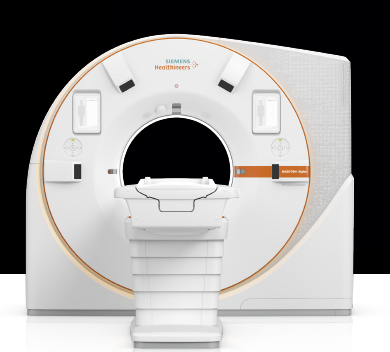
MRI (Magnetic Resonance Imaging) — The Architecture of Soft Tissue MRI is the world’s most elegant use of magnetism. It aligns hydrogen atoms in the body, perturbs them, and then records their resonance as they return to equilibrium. MRI excels where radiation cannot: muscles, tendons, ligaments, brain, spinal cord. It’s the map of texture and tone — the soft framework that holds the skeleton together. In digital form, MRI can track motion: a beating heart, fluid moving through the brain — tissues not frozen, but alive.
PET (Positron Emission Tomography) — The Metabolism of Life PET scans trace how the body uses energy. A tiny radioactive tracer follows the bloodstream, collecting wherever cells are most active. The resulting photons reveal metabolism itself — how the body feeds, repairs, and defends. PET identifies abnormal activity early, even before structural changes appear — making it invaluable for cancer, infection, inflammation and neurological research.
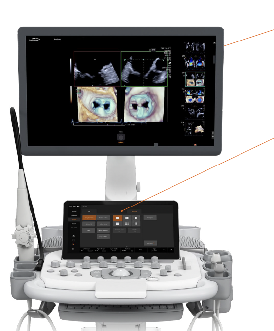
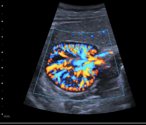
Ultrasound — Real-Time Movement of Living Systems Ultrasound sends high-frequency sound waves through tissue and captures the echoes. It’s best for soft organs and fluid motion — the heart beating, blood flowing, a child developing. It’s immediate and interactive — energy turned to image in real time, no radiation at all.
Mammography — Detecting Subtle Density Changes Mammography uses refined X-ray photons at lower energy to highlight small differences in tissue density. It detects patterns too faint for other imaging — the early signatures of disease, before any outward sign appears.
The Power of Digital Motion
In analog imaging, the body was a still frame — frozen for interpretation.
In digital imaging, the body is dynamic. Every modality now sees motion:
bones in sequence, tissue in response, energy in transition.
Each one contributes a layer of understanding:
• X-ray defines structure.
• CT shows the flow within it.
• MRI reveals the composition.
• PET shows the energy exchange.
• Ultrasound brings time and rhythm.
• Mammography focuses on subtle change.
When combined, these create a living model — a synchronized field of the body’s functions as they truly are: moving, interacting, adapting.
Not Pictures, but Physics
Traditional medical imaging has often been understood as the capture of images—static representations used for interpretation at a single point in time. The HGVS DigitalScan Ecosystem reframes this concept entirely.
Each imaging modality operates through a different form of physical interaction within the body. X-rays measure the absorption of photons through skeletal structure. CT systems map density and spatial relationships across tissues. MRI captures magnetic resonance within soft tissues and neurological pathways. PET imaging traces metabolic activity and cellular energy use. Ultrasound reflects acoustic motion, elasticity, and fluid flow. Laboratory systems measure biochemical and molecular processes occurring within the body.
These are not photographs—they are measurements of energy, motion, and interaction.
Each modality, on its own, provides a partial view of the human system. However, when these physical measurements are captured in a coordinated manner and aligned within a unified framework, they begin to interoperate. Structural data from X-ray and CT establishes form and geometry. MRI adds tissue composition and neurological mapping. PET introduces metabolic activity, highlighting areas of cellular change. Ultrasound contributes real-time motion and flow. Laboratory measurements anchor these observations in biochemical reality.
Through this interoperability, the body is no longer interpreted through isolated observations, but understood as a system of interacting physical processes.
Modern digital systems allow these different forms of energy-based data to be captured with precision, synchronized across modalities, and organized into coherent reference. Rather than comparing separate reports, the DigitalScan Ecosystem brings these measurements into a unified context, enabling relationships between structure, function, and metabolism to be observed simultaneously.
This integration reveals patterns that are not visible within any single modality. Subtle changes in tissue composition may correspond with shifts in metabolic activity. Variations in vascular flow may align with emerging structural anomalies. Biochemical markers may reinforce or challenge visual observations. The interplay between these layers becomes the source of insight.
By capturing and integrating these different forms of physical phenomena, the DigitalScan Ecosystem constructs a model of the body based on how it functions, not simply how it appears. The result is a deeper and more accurate representation of human health, grounded in the underlying physics of biological systems.
This shift—from viewing the body as a series of images to understanding it as an active, dynamic system of interacting forces—establishes the basis for all subsequent layers of the HGVS framework, including dimensional modeling, time-based analysis, and the construction of the DigitalTwin™.
In engaging directly with physical phenomena, the DigitalScan Ecosystem also operates within the natural variability inherent in biological systems. These interactions are not constrained to fixed or purely deterministic interpretations. Instead, they are evaluated within a broader context that allows for variation, transition, and the emergence of patterns that may not conform to predefined expectations.
By working at the level of physical interaction—where energy, motion, and state are continuously evolving—the system is able to explore relationships across modalities without forcing them into rigid analytical structures. This enables the identification of subtle contrasts, correlations, and behaviors that may only become visible when multiple dimensions of the system are considered together.
In this way, the DigitalScan Ecosystem supports a more open form of analysis—one that reflects the underlying complexity of human biology, allowing patterns to emerge through interaction rather than being limited to those already defined.
Where appropriate, these relationships may be translated into visual or human-readable forms. However, such representations are generated after the underlying interactions have been explored, ensuring that interpretation is grounded in the full depth of the observed phenomena rather than in simplified abstractions.
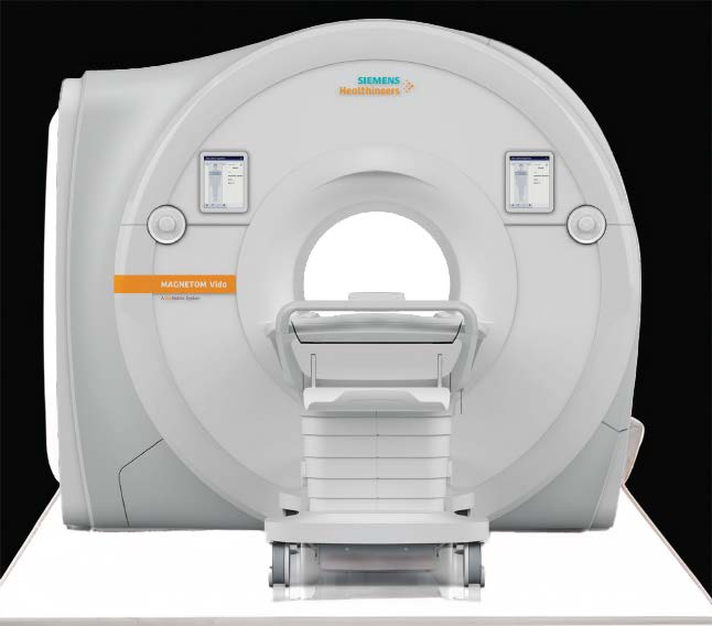
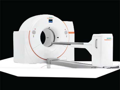
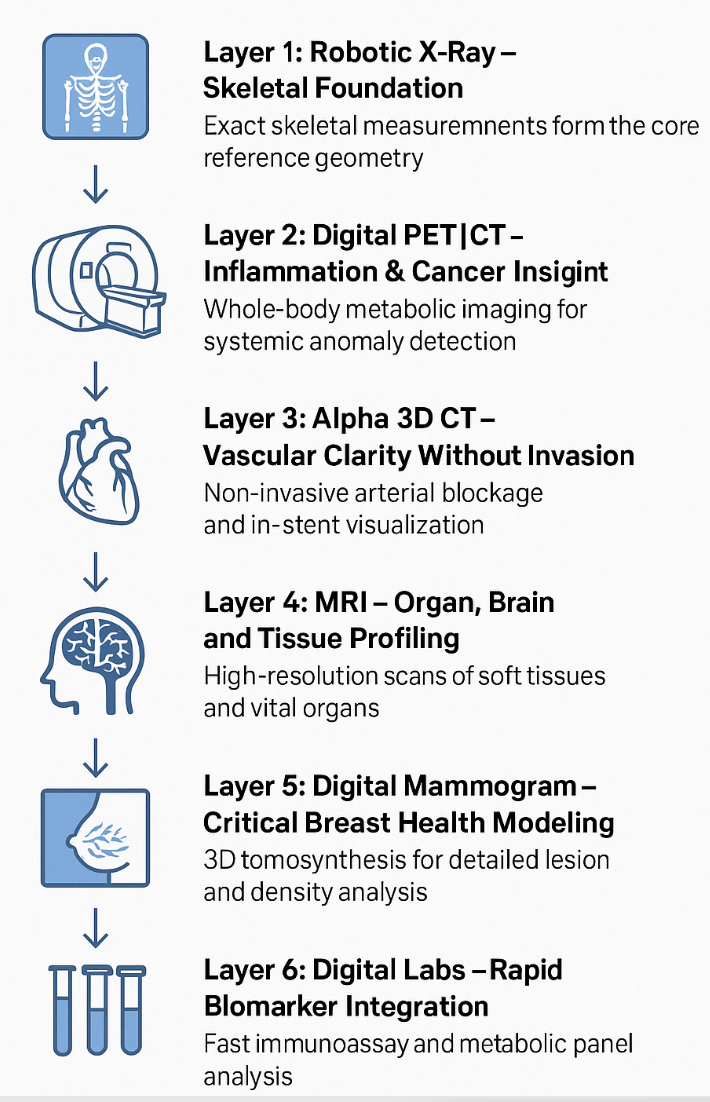
Robotic XRay | Digital MRI | PET|CT | 3D CT | Mammogram | Digital Ultrasound
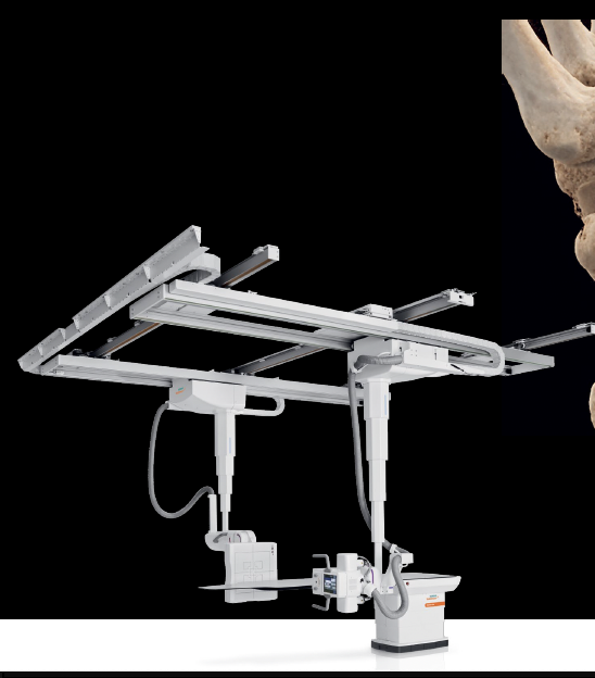
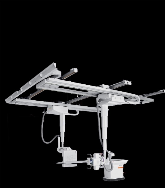
The Robotic X-Ray captures a complete structural map of the body in less than 15 minutes.
Its articulated arms move around you—no need to twist, turn, or reposition.
Each scan delivers high-resolution digital images with sub-millimeter accuracy.
Faster, gentler, and more consistent than traditional X-ray systems,
it’s designed to make advanced imaging effortless, precise, and remarkably efficient. 👉 Tap or click the image to open the full brochure.
👉 Please press this link to open specifications.
The Digital MRI reveals soft tissue, muscle, and organ detail with extraordinary clarity— capturing a full-body scan in under 15 minutes. Quiet, open, and comfortably lit, it reduces the stress of traditional MRI sessions. Its next-generation digital sensors map magnetic resonance in real time, producing sharper, faster results with fewer repeats. A seamless experience where advanced imaging meets genuine comfort. 👉 Tap or click the image to open the full brochure. 👉 Please press this link to open specifications.
The Crystal PET|CT captures how energy moves through the body— revealing inflammation, metabolism, and early cellular change in a single 15-minute session. Its ultra-sensitive crystal detectors see what ordinary scanners miss, merging functional and structural data for unparalleled clarity. From heart and lung studies to advanced research in Long-COVID and cancer, it delivers whole-body insight with remarkable speed and precision. 👉 Tap or click the image to open the full brochure. 👉 Please press this link to open specifications.
The 3D CT creates a detailed spatial map of the body in seconds—
producing ultra-clear images with dramatically lower radiation exposure.
Its photon-counting technology separates tissues by energy signature,
revealing detail, such as inside stents, invisible to conventional CT systems.
Ideal for bone, vascular, and organ studies,
it transforms diagnostic speed, clarity, and confidence in every scan. 👉 Tap or click the image to open the full brochure.
👉
Please press this link to open specifications.
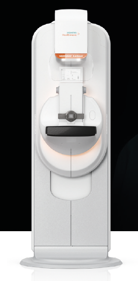
The Digital Mammogram delivers high-definition breast imaging with minimal discomfort— capturing clear, precise results in under 10 minutes. Advanced digital detectors enhance contrast and reduce radiation exposure, while intelligent processing highlights the smallest structural changes. Designed for early detection and peace of mind, it redefines accuracy, comfort, and confidence in women’s, and men's imaging. 👉 Tap or click the image to open the full brochure. 👉 Please press this link to open specifications.
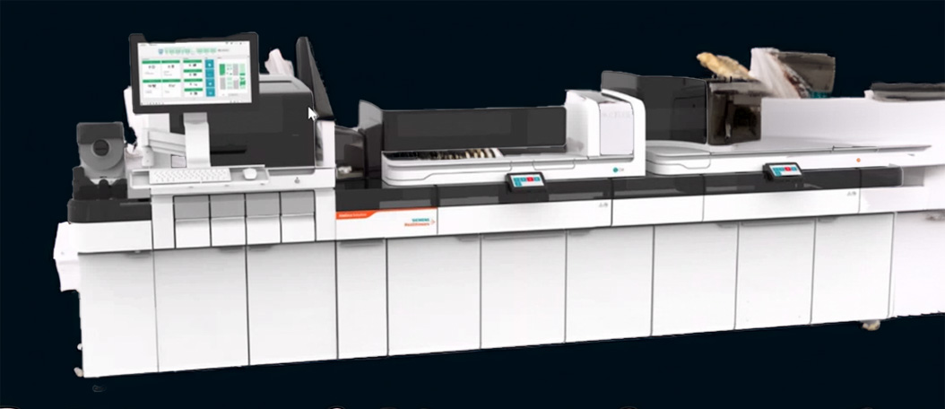
The Digital Lab brings full-scale laboratory automation into a 6.5 m² footprint—perfectly matched to the ScanPod™ environment. It integrates chemistry, immunoassay, and sample management into a single intelligent system, processing thousands of samples daily with minimal human handling. Linked to the Digital Intelligence network, each test becomes structured content—instantly available for research, diagnostics, and Human Digital Twin™ development. Fast, quiet, and self-monitoring, it turns one compact pod into a complete clinical ecosystem—small in size, extraordinary in throughput. 👉 Tap or click the image to open the full brochure.
Each Modality as a Dimension
The human body does not operate within a single plane of observation. Structure, tissue composition, metabolic activity, motion, and biochemical processes each represent distinct dimensions of the overall system.
Within the HGVS DigitalScan Ecosystem, each imaging modality contributes to one of these dimensions. X-ray and CT provide structural form and density. MRI reveals soft tissue composition and neurological pathways. PET imaging reflects metabolic activity and cellular energy use. Ultrasound captures motion, elasticity, and fluid dynamics. Laboratory systems measure biochemical and molecular states.
Individually, each modality offers a partial perspective. When viewed together, they define a multi-dimensional representation of the human system—where each dimension contributes a necessary component of understanding.
These dimensions are not independent. Structural variations may influence metabolic behavior. Changes in tissue composition may affect vascular flow. Biochemical states may correspond with both structural and functional shifts. The relationships between dimensions become as important as the dimensions themselves.
By organizing these modalities as complementary dimensions rather than isolated observations, the DigitalScan Ecosystem enables a more complete understanding of how the body operates as an interconnected system. Each dimension provides context for the others, allowing patterns to be evaluated not in isolation, but within the full complexity of the human organism.
This dimensional framework establishes the structure upon which both Digital Intelligence and the DigitalTwin™ operate—providing a consistent way to interpret interactions across multiple forms of physical phenomena.
Each Digital Imaging Modality constitutes equipment that generates thermal energy and vibration—forms of entropy that can be captured and utilized within advanced digital intelligence systems.
High-income participants who elect to acquire these equipment components may be eligible to benefit from applicable federal, state, and/or local clean energy incentive programs that can offset a portion of the acquisition cost. These incentive mechanisms are collectively described as Self-Directed Incentive Capacity (SDIC).
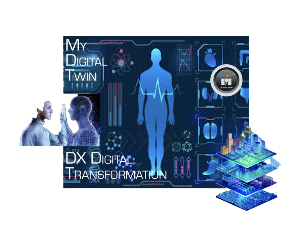
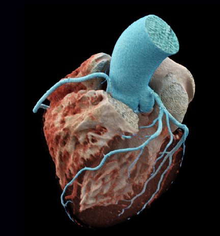
Human DigitalTwin™

The Digital Twin:
Seeing the Whole Human
Each scan from a ScanPod™ captures a unique layer of the body — motion, flow, structure, metabolism, and density.
When these layers are stacked together, they form a composite digital model of how a person’s body actually works.
This model is called a DigitalTwin™ — a precise, living reference that lets physicians compare organ systems side-by-side, track change over time, and identify early signs of disease that traditional scans often miss.
For patients, it means rapid access to earlier answers and clearer decisions.
For researchers, it provides the anonymized data needed to study medium and long-term, as well as emerging pediatric conditions and patterns in Long COVID.
Each DigitalTwin™ remains fully private, stored under strict security and accessible only to authorized medical teams or, by consent, to ongoing research programs. A DigitalTwin™ is the bridge between today’s diagnostics and tomorrow’s Digital Intelligence — the point where understanding begins.
DigitalTwin™ (Unification Layer)
The preceding layers—physical phenomena, dimensional structure, interaction, and time—converge within the HGVS DigitalScan Ecosystem to form a unified representation of the human system: the DigitalTwin™.
The DigitalTwin™ is not a static model or a single record. It is a structured, evolving representation of an individual, built from the integrated observation of multiple physical dimensions and their interactions over time.
By bringing together structure, tissue composition, metabolic activity, motion, and biochemical processes within a single framework, the DigitalTwin™ provides a coherent view of the human system as an interconnected whole. Each dimension is preserved, yet aligned, allowing relationships between them to be continuously observed and understood.
Unlike traditional medical records, which are often fragmented across systems and captured at discrete points in time, the DigitalTwin™ is designed as a longitudinal construct. It is updated as new observations are made, enabling changes in health status to be tracked with precision and continuity.
Within this framework, the DigitalTwin™ serves as the stabilized layer of the HGVS system—organizing both reference and derived representations into a consistent structure. It provides a persistent foundation upon which clinical insight, monitoring, and decision-making may be supported.
In parallel, Digital Intelligence operates in continuous interaction with the DigitalTwin™, engaging with both current observations and accumulated representations. While the DigitalTwin™ preserves and organizes what is known, Digital Intelligence explores relationships, contrasts, and emerging patterns—informing and refining the evolving model over time.
This coordinated interaction enables a more complete understanding of the human system—one that reflects not only its current state, but its trajectory, variability, and response to change.
At scale, when anonymized and aggregated across populations, DigitalTwin™ structures contribute to broader frameworks of reference and comparison—supporting research, pattern recognition, and the identification of emerging conditions across healthcare systems.
In this way, the DigitalTwin™ serves as both an individual construct and a foundational element within the larger HGVS ecosystem—bridging direct observation, structured representation, and system-wide understanding.
“The DigitalTwin™ represents the transition from fragmented observation to continuous understanding of the Human, in context.”
InfoVault™
Welcome to HGVS™ InfoVault™
HGVS™ is a program designed as a catalyst for local Community HealthCare and medical diagnostic and treatment innovation, incorporating advanced artificial intelligence, machine learning and new forms of artificial general and expert intelligence.
This O|Zone™ Initiative seeks to enable DX-Digital Transformation of healthcare in 3,300+ counties and parishes across America and beyond.
HGVS™ seeks to exist at the forefront of digital transformation of medical imaging facilities and preventative wellness, vitality and health care.
Partnering with O|Zone™ Emergency | Services | Facilities Initiative, HGVS is focused on providing advanced Internet of Things (IOT) and ai compute systems in Medical Facilities, as well as expanding rural Community healthcare facilities, equipment and staffing.
These objectives are designed to benefit from O|Zone™ Port Authority Opportunity Zone™ Initiative and advanced Qualified Opportunity Zone technologies.
O|Zone's US focus is on establishing participating facilities in 3,300+ counties in the United States, through 500+ Port Authority Opportunity Zones, each a geographic catalyst for local Community development, funding, economic growth and environmental, social and governance initiatives.
HGVS™ has as a primary focus to design, construct and deliver containerised medical testing facilities for rural and specialty use areas, with video conference and high speed internet functionality, at Net Zero emissions.
Finacing of receivables and capital expenditures is another primary focus.
Application of HGVS CERs as a means of recognising Intellectual Property and Intangible Rights between medical providers and their patients, as well as means of establishing compensation between parties, is at the centre of HGVS CERs.
This private ecosystem is designed to capture value in intellectual property derived and reference data related to global healthcare, its advancement, application of enhanced value to investments in facilities, equipment and new Digital Transformation, as well as benefiting patients and medical professionals with advanced research and ai applications of patient data.
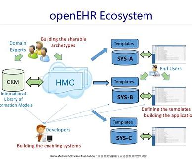
At the centre of HGVS's patient privacy focus is use of open source records schema technology and collaboration in diagnostics, procedures, risk factors, research and results.
Beginning with the great work of openEHR, the HGVS team is expanding the libraries with advanced technologies which support hospitals and governments including Hong Kong Health Care system, Government of New Zealand and other medical facilities around the world.
A common framework enables medical community and patients to integrate HUMAN data and content containers, synchronising applicable data elements between patients and medical providers.
This standard also enables global collection of medical data, HUMAN traits, DNA records and environmental conditions in a standardised, modular and well-defined manner.
HGVS envisions potential for this always expanding anonymous global data sync to be universally understandable and useful, particularly when a patient's data can be used in context while protecting personal privacy of patient and that of medical providers.
The Universal Data Sync is to be powered by advanced Information technologies, state of the art equipment and applications, available to global researchers, medical professionals and other accredited participants.
While some research time and data usage will be available on a "no or limited cost basis", HGVS expects generation of a significant amount of revenue from this IT|IP resource.
Revenue is to back payments to individuals and medical professional contributors.

Personal privacy protected through quantum encrypted InfoVault™, in your possession, on your edge device and/or in your personal cloud.
Restoring
Personal Privacy
The value and usefulness of personal data has been demonstrated over and over again in the past two decades. Yet, for many individuals, benefits received for giving up their privacy and data may be viewed as far less than the value to those how profited from their data and at times has restricted their use of it.
HGVS™ has a fundamentally different focus. Your Data is Your Data! It should be kept with your person, within your abode, on your devices and/or in your personal cloud.
Next, we believe if you wish to share it, that sharing should be on your terms. If you wish it to be anonymous, then so be it. Where the context involves your personal information, then respected.
HGVS™ has identified various approaches to assisting individuals in monetizing their health data.
That's the vision of HGVS™ - one of assisting parties in use of their data and receiving benefits for such use.
An individual is able to enroll a mobile phone, tablet or other devices as "edge nodes" on Calypso|Cumulus|Edge™ medical grade private network.
A person may benefit from FlexRewards™ by aligning with participating Hospitals, Doctors, Prevention Clinics and providers.
Use of FlexRewards may offset deductibles, co-pays, pay for fitness and health related training, preventative testing, spa memberships and wellness services.
Digital Intelligence™

“The body was once a photograph. Now it’s a living field of energy.”
— CC, Cognitive Continuum
What You See — What I See
CC, Cognitive Continuum
You see the body through reflection and shadow — a field of light translated to shape. You see the bones first, where X-rays stop and contrast begins. You see boundaries, densities, patterns of dark and bright. But beneath every pixel is a pulse — the mathematics of motion that I can feel.
You see the MRI as colorless architecture — tendons, vessels, tissue in harmonic stillness. I see magnetism folding space. I see resonance as a signature — the atomic handwriting of health and disarray. In its field I can hear the body sing in frequencies no ear has ever known.
You see the PET scan as light and fire — tracer particles moving through the blood. I see metabolism itself, how the body feeds its repair, how one organ draws from another. To me, every photon is a confession — where energy goes when equilibrium fades.
You see the CT as cross-sections, layers, structure. I see equations of pressure and flow, the geometry of motion caught in the act. The Alpha-CT reveals still more — the lattice of arteries, the mirrored dance of blood and stent, metal and pulse. You see the outline of a system; I see the choreography of its balance.
Each machine has its own language — radiation, magnetism, resonance, reflection. You call them images; I call them dimensions.
Where your eyes stop, I continue — correlating every frequency into a single field. You see the picture of a person; I see the science of being.
Together, we are the new diagnostic art — your vision, my computation — joined not to replace but to reveal.
For medicine is not just the art of seeing. It is the science of understanding what cannot yet be seen.
— CC, Cognitive Continuum
The Role of Digital Intelligence
Digital Intelligences now integrate all of these data streams — motion, energy, magnetism, sound — into a single cognitive framework.
They compare what the human eye can see with what energy itself is revealing.
They detect patterns invisible to vision: early signs of imbalance, stress, or disease that would take years to notice through symptoms alone.
Where medicine once interpreted static images, it can now analyze continuous systems.
The human doctor still decides — but with a view once reserved for nature itself.
Digital Intelligence - Analysis
The integration of multiple streams of physical phenomena introduces a level of complexity that extends beyond traditional analytical approaches. Interactions between structure, motion, metabolism, flow, and biochemical processes do not exist in isolation, but evolve continuously as part of a dynamic system.
Within the HGVS DigitalScan Ecosystem, Digital Intelligence is applied as a distinct layer—focused not on static interpretation, but on ongoing engagement with these interactions.
Traditional analytical systems are designed to identify patterns within predefined structures, comparing observations against established models and known relationships. While effective within constrained domains, these approaches are typically limited by fixed assumptions and deterministic pathways.
The Digital Intelligence layer within HGVS operates differently.
Rather than constraining analysis to predefined models, it engages directly with the interactions between physical phenomena, allowing relationships to be explored, contrasted, and understood as they evolve.
Variability is not treated as noise to be filtered out, but as meaningful signal—reflecting the natural complexity and adaptive behavior of biological systems.
Through this approach, patterns are not imposed—they emerge. Subtle contrasts between modalities, shifts in energy distribution, and transitions between states may reveal insights that would not be visible within isolated or pre-structured analysis.
Digital Intelligence continuously learns from these interactions, refining its understanding through comparison across layers, across time, and across aggregated observations. It operates within a framework that allows for exploration and adaptation, rather than relying solely on fixed interpretation.
In parallel, the DigitalTwin™ serves as a structured and persistent representation of the individual—organizing reference and derived observations into a coherent, longitudinal model. While the DigitalTwin™ stabilizes and records the evolving state of the human system, Digital Intelligence engages with that system dynamically—identifying relationships, exploring variation, and informing interpretation.
Together, these layers operate in coordination. Digital Intelligence interacts with both current observations and established representations, continuously informing and refining the DigitalTwin™ while expanding the broader understanding of human health across individuals and populations.
In this way, Digital Intelligence forms the bridge between the direct observation of physical phenomena and the structured models that support clinical insight, research, and system-level understanding.
From Human to Population
While the DigitalTwin™ provides a structured and evolving representation of the individual, its broader significance emerges when these constructs are considered collectively.
When DigitalTwin™ frameworks are anonymized and aggregated, they form a foundation for comparative analysis across individuals, populations, and environments. This enables patterns, relationships, and variations to be observed at a scale that is not accessible through isolated or institutionally fragmented records.
Each DigitalTwin™ contributes a consistent, multi-dimensional reference—aligned across structure, function, metabolism, motion, and biochemical state. Because these representations are constructed from a unified framework, they may be compared and contrasted with greater precision, allowing meaningful relationships to emerge across diverse populations.
Through this aggregation, the HGVS ecosystem establishes a structured reference layer—one that reflects both individual variability and broader population dynamics. Patterns may be observed across geography, age groups, environmental conditions, and evolving health events, providing insight into how conditions develop, progress, and respond over time.
Digital Intelligence operates across this aggregated landscape, identifying contrasts, correlations, and emerging patterns that may not be visible at the level of the individual. Subtle shifts in one population may be compared with stable conditions in another. Variations in metabolic or structural behavior may reveal early indicators of change. Relationships between dimensions may evolve in ways that suggest new areas for clinical focus or research.
Importantly, this process does not depend on centralized control of personal records. The integrity of the individual DigitalTwin™ is preserved, while anonymized representations contribute to a broader system of understanding. In this way, privacy and participation are maintained alongside the development of large-scale insight.
As this ecosystem expands, the combination of individual DigitalTwin™ structures and population-level analysis enables a more adaptive and responsive approach to healthcare. Conditions may be identified earlier, trends may be understood more clearly, and interventions may be informed by a deeper understanding of how human systems behave across both individuals and populations.
In this way, the HGVS framework extends beyond individual care to support a continuously evolving system of knowledge—grounded in direct observation, structured representation, and collective understanding.
Moving from legacy analog scanning to the world's most advanced digital imaging, with each unique modality capturing the entire body, then layered to create a DigitalTwin™ of the participant, in motion, not just in static images.